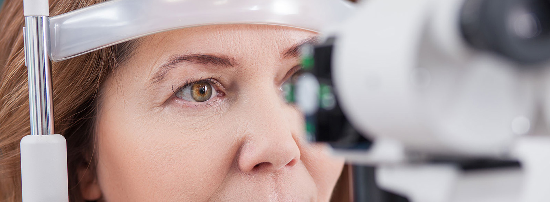
Diabetes affects more than blood sugar — it can gradually change the structure and function of the eyes. Over time, high glucose levels can damage the tiny blood vessels that nourish the retina, leading to conditions that may quietly reduce vision long before symptoms appear. Because these changes often develop without pain or noticeable warning signs, regular, comprehensive eye exams are the single most effective way to catch problems early.
Early detection dramatically improves the odds of preserving sight. Identifying retinal changes at a mild stage opens the door to monitoring and interventions that can slow or stop progression. That’s why eye care professionals recommend a baseline dilated exam at diagnosis and at least yearly follow-ups for most people with diabetes — more frequently if changes are detected or if systemic control fluctuates.
An annual diabetic eye exam is also an opportunity to coordinate care. Eye findings can reflect how well diabetes is being managed overall, and timely communication between your optometrist, primary care provider, and endocrinologist helps align treatments that protect both general health and vision. Think of the exam as part of a broader diabetes-management team effort, not just a single appointment focused on glasses.
A diabetic eye exam goes beyond a standard vision test. After checking your visual acuity, the doctor typically dilates your pupils to get a wide view of the retina. Dilation allows the clinician to examine the retinal surface and optic nerve for signs of bleeding, swelling, new abnormal blood vessels, or other structural changes associated with diabetic eye disease.
Modern imaging tools—such as optical coherence tomography (OCT) and retinal photography—are often used to document the retina’s condition in detail. OCT provides cross-sectional images that reveal swelling or thickening at the macula, while retinal photos create a permanent record clinicians can compare over time. These technologies increase diagnostic precision and make it easier to recognize subtle changes between visits.
Intraocular pressure testing and an assessment of the anterior eye are also important components. People with diabetes have a higher risk of glaucoma and cataract formation, and screening for these conditions helps the doctor form a complete picture of eye health. Together, these tests build a thorough, reliable evaluation that supports early detection and tailored care plans.
Although many diabetic eye changes are silent, certain symptoms should prompt immediate attention. Sudden blurring, the sudden appearance of floaters or cobwebs, flashes of light, or a shadow or curtain across any part of your vision are all red flags that warrant an urgent evaluation. These signs can indicate bleeding inside the eye or retinal detachment, both of which require prompt assessment.
Gradual symptoms can also signal trouble: a slow decline in reading sharpness, fluctuating vision throughout the day, or persistent distortion where straight lines appear wavy should not be ignored. Because some symptoms may overlap with other eye conditions, a comprehensive exam is the only reliable way to determine the cause and next steps.
If you have diabetes and experience any new visual changes, don’t wait for your next scheduled visit. Quick reporting and timely assessment improve the chances of effective treatment and preservation of sight, so contact your eye care provider as soon as you notice unusual changes.
Protecting vision starts with controlling the systemic factors that drive diabetic eye disease. Consistent blood sugar management reduces the stress on retinal blood vessels, while controlling blood pressure and cholesterol lowers the risk of vascular complications that affect the eye. These elements work together: better metabolic control reduces the likelihood of progressive eye damage and improves outcomes if treatment is needed.
Lifestyle measures complement medical therapy. Regular physical activity, a balanced diet, and smoking cessation all contribute to healthier circulation and a lower overall risk profile. Routine monitoring of glycemic control, blood pressure, and lipid levels — along with adherence to prescribed medications — supports both general health and long-term vision preservation.
Coordination between your eye care team and primary medical providers is essential. Sharing exam findings and imaging results helps clinicians adjust systemic treatment plans when eye changes suggest a need for tighter control. In this way, eye exams serve not only to protect vision but also as a barometer of overall vascular health.
When diabetic eye disease is identified, the approach depends on the type and severity of the changes. Some early alterations can be managed with careful observation and more frequent monitoring, while more advanced findings may require medical or surgical treatments aimed at stabilizing or improving vision. The goal of any intervention is to prevent further loss and maximize quality of sight.
Common medical treatments include targeted injections that reduce swelling or inhibit abnormal vessel growth, and laser therapies that seal leaking vessels or reduce the risk of further bleeding. In cases where bleeding or scar tissue threatens the retina, surgical options such as vitrectomy are considered. The choice of treatment is individualized, based on imaging, clinical findings, and patient-specific considerations.
Follow-up schedules are tailored to each person’s situation. Some patients return every few months during active treatment phases, while others may be monitored annually when disease is stable. Consistent follow-up with retinal imaging helps measure response to therapy and guides adjustments to the plan. Our team stays focused on timely, evidence-based care and clear communication about what to expect during treatment and recovery.
At Vision World of Copiague, our clinicians combine current diagnostic technology with collaborative care strategies to detect and manage diabetic eye disease early. We emphasize monitoring, patient education, and coordination with your medical providers so that treatment decisions reflect both ocular needs and overall health goals.
Keeping vision healthy when you have diabetes requires vigilance, timely exams, and a partnership between you and your care team. If you have questions about diabetic eye exams or would like more information about what to expect, please contact us for more information.
A diabetic eye exam is a comprehensive evaluation that focuses on detecting early signs of diabetic eye disease and related complications. It typically includes a dilated retinal exam that allows the clinician to inspect blood vessels, the macula and the optic nerve for subtle changes. The exam often combines visual acuity testing, retinal imaging and other measurements to identify problems that may not yet be causing symptoms.
The primary goal of a diabetic eye exam is early detection so appropriate interventions can begin before significant vision loss occurs. Examiners look specifically for diabetic retinopathy, diabetic macular edema, cataract development and glaucoma. Advanced imaging such as optical coherence tomography or retinal photography may be used to document and track subtle changes over time.
People with any form of diabetes should have regular diabetic eye exams because chronically elevated blood sugar can affect the retina, lens and optic nerve. In general, a comprehensive dilated eye exam at least once a year is recommended, with more frequent follow-up when retinopathy or other complications are present. Specific timing may differ for children, pregnant patients and those with long-standing disease, so your eye doctor will tailor recommendations to your situation.
Pregnant people with diabetes and those planning pregnancy should have a baseline exam and close monitoring throughout pregnancy because hormonal and metabolic changes can accelerate retinal changes. Individuals with hypertension, kidney disease or poor glycemic control may also need more frequent exams to protect vision. Discussing your overall health with both your eye doctor and primary care provider helps coordinate optimal care.
Most people with diabetes should have a comprehensive dilated eye exam at least once every 12 months, but individual needs vary based on disease duration and stability. If retinal changes are detected, the eye care team may recommend checkups every three to six months or even more frequently to monitor progression. Prompt follow-up allows clinicians to apply treatments at stages when they are most effective.
Vision World of Copiague works with patients to develop a personalized surveillance plan that balances regular monitoring with practical scheduling. Factors that influence frequency include blood sugar control, presence of retinopathy or macular edema, pregnancy and concurrent eye conditions such as glaucoma or cataract. Be proactive about scheduling as recommended so treatments, when needed, can begin promptly.
A diabetic eye exam usually begins with a review of medical history and current medications followed by visual acuity testing to assess clarity of vision. Pupil dilation is commonly performed so the doctor can examine the retina, macula and optic nerve for signs of damage or leaking blood vessels. The visit may also include intraocular pressure measurement and assessment of the front structures of the eye.
Clinicians often use imaging tools such as optical coherence tomography and retinal photography to document retinal thickness and blood vessel changes. These tests are painless and help detect subtle abnormalities that are not apparent on routine examination alone. Depending on testing and dilation, the entire visit generally takes 30 to 60 minutes.
Common symptoms include blurred or fluctuating vision, dark spots or floaters, difficulty reading and distortion in central vision. Some people notice reduced contrast sensitivity or that colors appear faded or washed out. The exact symptoms depend on whether the macula, retina or other eye structures are affected.
Early stages of diabetic retinopathy are often asymptomatic, so waiting for symptoms can delay treatment. Any sudden changes in vision, new floaters, flashes of light or a shadow across the field of vision require immediate attention. Regular exams allow the eye care team to find problems before they cause irreversible damage.
Chronically elevated blood sugar can damage the small blood vessels in the retina, producing diabetic retinopathy where vessels leak or close off and create areas of retinal ischemia. When these leaks occur in the macula, they cause diabetic macular edema that directly impairs central vision and reading ability. Diabetes also increases the risk of cataract formation by altering lens proteins and may contribute to higher intraocular pressure associated with glaucoma.
These processes can occur together, so a patient may experience multiple eye conditions at once, making comprehensive assessment essential. Controlling systemic risk factors and close ophthalmic monitoring are key strategies to slow progression and preserve vision. Early diagnosis and coordinated care between eye specialists and medical providers improve long-term outcomes.
Treatments are available that can slow disease progression, reduce vision-threatening swelling and address abnormal blood vessel growth, although reversal of advanced damage is limited. Common interventions include intravitreal injections to reduce macular edema, laser therapy to seal leaking vessels and vitrectomy surgery for severe bleeding or tractional retinal detachment. Tight control of blood sugar, blood pressure and cholesterol supports eye treatments and lowers the risk of further damage.
Vision World of Copiague coordinates with medical providers to ensure patients receive prompt evaluation and timely referral to retinal specialists when advanced therapy is needed. Regular monitoring enables these treatments to be applied at stages when they are most effective in preserving vision. Early and consistent follow-up improves the likelihood of maintaining functional sight.
Bring a list of current medications, recent blood sugar or A1C results and any documentation of prior eye care or retinal images if available. Also bring your current glasses or contact lens information so visual acuity can be assessed accurately. Providing a concise medical history helps the clinician understand systemic risks that influence eye health.
Because pupil dilation is often required, plan for temporary blurred near vision and light sensitivity and arrange transportation if needed. If you experience significant vision changes before your scheduled visit, contact your eye care team for an earlier appointment. If you use insulin or other diabetes medications, discuss timing and food intake to minimize hypoglycemia during the visit.
Optical coherence tomography (OCT) is a noninvasive, high-resolution scan that measures retinal thickness and detects macular edema. Wide-field and standard retinal photography document blood vessel changes and make it easier to track progression over time. Fluorescein angiography may be used selectively to study blood flow in the retina and identify leaking or nonperfused areas.
These imaging modalities complement the dilated clinical exam and often reveal subtle disease before symptoms appear, guiding timely treatment decisions. Imaging results are stored as part of your record so clinicians can compare scans over time and measure response to therapy. Your eye doctor will recommend the most appropriate tests based on clinical findings and disease severity.
Diabetic eye disease can develop silently for years, and early changes often do not affect central vision until significant damage has occurred. Regular examinations detect these early signs so that interventions can be started before vision loss becomes permanent. Detection also allows the care team to monitor treatment effectiveness and adjust management promptly.
Staying current with eye exams is one of the most effective steps a person with diabetes can take to protect long-term vision, alongside systemic health management. If you have not had a dilated retinal exam within the past year, contact your eye care provider to schedule one and discuss personalized follow-up. Early partnership between patients and clinicians improves outcomes and quality of life.
Quick Links
Contact Us